
- 4 February 2023
- Comment: 0
- Treatments
The aneurysm that occurs in the vessel due to the weakness of the muscle layer on the wall of the brain vessels is called a cerebral aneurysm. This aneurysm causes thinning and weakening of the vessel wall. Brain hemorrhage occurs as a result of the rupture of the vein from this place where it is weakened. Brain aneurysms can occur in people of any age, but most are in people between the ages of 35 and 60. Women are slightly less likely to have an aneurysm than men.
Aneurysms are formed at the points where the vessel wall is congenitally weak, usually at the points where the vessel divides into smaller branches. Due to the intravascular pressure (blood pressure) at the point where the vessel wall is weak, the vessel wall bulges outward from the weak point in each heartbeat and bubbles are formed. The bubble wall bursts as soon as it cannot withstand the pressure; the explosion either occurs spontaneously or occurs with effort.
The result of long-term follow-up of aneurysms detected in brain vessels is the risk of bleeding at an increasing rate of 0.2-3% every year. Especially in young people, when it is considered that the patient has a long life ahead and this bleeding rate is added every year, the probability of bleeding increases even more. Therefore, treatment of this aneurysm is recommended for people who are found to have an aneurysm.
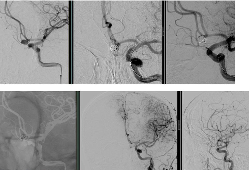
Interventional neuroradiologists perform this procedure in the angiography unit while the patient is under general anesthesia. Without making a surgical incision, the artery in the groin is entered with a needle and a millimeter-sized plastic tube called a catheter is inserted into the artery. This catheter is advanced to the neck veins leading to the brain. A microcatheter is advanced through this catheter into the aneurysm. The aneurysm is blocked with platinum coils (coils) placed through the microcatheter and blood is prevented from entering it. In some cases, a stent or balloon can also be used to assist the coiling procedure and protect normal vessels adjacent to the aneurysm. As a result, by means of the embolization procedure, brain aneurysms are treated without the need for open surgery and the risk of bleeding is prevented.
Example case images:
Other cerebrovascular diseases treated with endovascular (angiography) method
- Brain hemorrhages (Due to bubble or glomerulus)
- Aneurysm (bubble) and AVM (glomerulus) that has not yet bled
- Leaks between arteries and veins (AVF)
- Treatment of vasospasm, which is frequently encountered after cerebral hemorrhage
- Fistula (abnormal blood passage between vessels)
- Adjunctive treatment for brain and spinal cord tumors
- Vascular stenosis
- Stroke
- In the treatment of childhood eye tumors